

Ultrasound
An ultrasound examination is a noninvasive medical test that helps diagnose and treat medical conditions. Ultrasounds capture the size, structure and shape of parts of the body that cannot be seen using conventional x-rays. An ultrasound exam is crucial in detecting changes or complications in organs, tissues, and vessels or to detect abnormal masses, such as tumors, gallstones, kidney stones, or liver disease by exposing specific body parts to high-frequency sound waves to produce pictures.
Ultrasound has been used by sonographers and radiologists (physicians who specialize in ultrasound) to image the human body for over 50 years, becoming one of the most widely used tools in modern medicine. Since there is no radiation involved, ultrasound is a safe test to examine pregnant women, evaluate the baby’s growth and determine due dates.
How do I prepare for the exam?
Preparation depends on the type of ultrasound you are having:
Abdominal ultrasound: You may be asked to fast (not eat or drink) for several hours before the exam.
Pelvic ultrasound: You may need to arrive with a full bladder for better imaging.
Other ultrasounds (breast, thyroid, vascular, musculoskeletal, etc.): Usually require no special preparation.
Our team will provide specific instructions when you schedule your appointment to make sure you are fully prepared.
What should I expect during the exam?
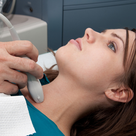
You will be asked to lie down comfortably on an exam table.
A small amount of clear gel will be applied to the skin over the area being studied. This helps transmit the sound waves.
The technologist will gently move a handheld device (called a transducer) over the skin to capture images.
The exam is safe, non-invasive, and painless.
Once complete, the images will be reviewed by a radiologist, and a detailed report will be sent to your doctor.