Tag Archives: Screening and Prevention

A preliminary procedure, such as a test or examination, to detect the most characteristic sign or signs of a disorder that may require further investigation, and the steps you can take to avoid unpleasant medical consequences.

CDI Miami | Tuesday June 21, 2016

The ALARA Principal

Intrinsic to working in radiology is working with radioactive materials. Although we will always be exposed to some level of radiation, we use the ALARA principal (As Low As Reasonably Achievable) to keep exposure to a minimum.


This principle assumes that there is no threshold for inducing “biological effects” and therefore any dose of radiation carries with it some risk. In other words, there is no level of “safe” radiation.


Using the strategies of Time, Distance, and Shielding, we can keep radioactive exposure as low as reasonably achievable. This maximizes efficacy for your treatment and keeps everyone safe, especially our patients. Below is a list of techniques we use to minimize radiation exposure:


alara principle

    1. Safe delivery of radiopharmaceuticals – When the syringe is being held with the plunger or needle, we make sure that it is managed with the utmost precaution, and, we take advantage of remote injectors whenever possible.


  • Transportation of Patients – How close to the back of the wheelchair are we standing when transporting a patient? Do we maintain appropriate distance when we are walking them out of the department or when we enter the scan room?


  • Cleaning up spills (including patient blood or body fluids) – This relates to how quickly we isolate and remove fluids that could contaminate the scan room, or mitigate the effect of the treatment.


  • Holding / Lifting Patients – We pay particular attention to where we stand in relation to our patients, especially if we have our radiation badge on.


  • Radioactive Tracers – When using these in treatment, we make sure that the all the precautions are made, so that we can have a safe and efficient treatment.


  • During PET/CT/MRI Scans – We drape our patient’s bodies with a radioactive shield, isolating the exposure to only the areas that need treatment.


These techniques are just a tiny sample of the ways we consider your wellbeing during our examinations. For diagnostic imaging services, including PET/CT Scans, MRIs, and Mammograms contact the Center for Diagnostic Imaging in Miami.


CDI Miami | Wednesday June 1, 2016

News: Chemo, Radiation, Surgery Combo Boosts Survival for Pancreatic Cancer Group

Recent Mayo Clinic research suggests that many pancreatic cancer patients whose tumors have grown around the pancreas to encompass critical blood vessels are candidate for surgery, even though conventional wisdom has suggested otherwise. The organization has been fine-tuning a protocol to treat this group – which accounts for roughly one-third of pancreatic cancer patients – and in two studies found survival now stretching into years.
“We’re definitely seeing a revolution,” said Mark Truty, M.D., a gastrointestinal surgical oncologist at Mayo Clinic in Rochester, Minn., who is first author of one abstract and senior author of the other. “A lot of this has to do with better chemotherapy drugs and use of what we call multimodal therapy: chemotherapy, radiation and then an aggressive operation. Now we can potentially offer these therapies to patients who previously were told they had no options.”

pancreatic cancer news

About 50,000 people are diagnosed with pancreatic cancer each year in the United States. Historically, only about 7 percent of pancreatic cancer patients have lived at least five years after diagnosis.

Because the cancer tends to spread before symptoms appear, it is found early enough to make surgery a clear-cut option in only about 15 percent of patients. In about half of patients, the cancer has spread throughout the body by the time it is diagnosed, ruling out surgery.

In one-third of patients, cancer hasn’t spread through the body, but has grown around veins and arteries in and around the pancreas. For decades, surgery was considered too risky and ineffective to be performed in most of those patients. The Mayo studies chronicle a transformation in treatment for these patients.

In the study presented at the Society for Surgery of the Alimentary Tract annual meeting, researchers analyzed surgical outcomes for the past 25 years among such stage 3 patients who had surgery requiring removal and reconstruction of arteries. They found that most of the operations on this group were performed in the past five years, since the advent of improved chemotherapy and radiation.

Although these surgeries carry more risk than operations not requiring removal and reconstruction of arteries, there appeared to be a significant long-term survival advantage in patients treated with chemotherapy and radiation followed by such aggressive operations. Those who had surgery without chemotherapy or radiation first didn’t do well long-term, while patients who had chemotherapy and/or radiation before surgery did significantly better long-term, the researchers found. Looking at short-term outcomes, they discovered that complication rates have decreased over time.

“All in all, it shows that these patients, who would typically not be offered an operation, can have good short-term and long-term results with the appropriate protocol and treatment sequence,” Truty said.

In the study presented at the Pancreas Club meeting, researchers analyzed modern surgical outcomes for stage 3 patients whose tumors involved blood vessels and who had a specific protocol of chemotherapy, radiation and aggressive surgery.

Eighty patients have now gone through the Mayo protocol with data available for review. The study found that the median survival time after patients complete the protocol is approaching four years, about four times that of patients who do not have surgery. The patients who do even better than that include:

  • -Those who receive more chemotherapy before surgery;
  • -People who have a particular tumor marker known as CA 19-9 that returns to normal after chemotherapy; and
  • -Those whose tumors, when analyzed after removal, are found to have only minimal cancer left.

The study also found that in a majority of patients, computed tomography (CT) scans before surgery showed that their tumors didn’t shrink after chemotherapy. However, when the tumors were removed, it turned out most of the cancer was dead.

“We’re hoping that data from this analysis will now spread to the rest of the country, and now people will have a road map for how to treat these patients and how to choose which patients will benefit from such complex operations,” Truty said. He hopes patients feel a sense of optimism, that there are options.

“Not everyone wants to sign up for these big operations or these long protocols of chemotherapy and radiation. But they have the options available to them to make that educated decision about whether this is something that would benefit them,” Truty said. “We’re offering an additional bit of hope for a pretty substantial number of patients who had previously been ignored.”

Originally published by Imaging Technology News.


CDI Miami | Tuesday May 17, 2016

How to survive an MRI or CAT scan if you’re claustrophobic

For some people, just thinking about an MRI is enough to provoke anxiety. For the approximately 9 percent of people diagnosed with claustrophobia, or a fear of enclosed spaces, “anxiety” is an understatement. Even for those without a clinical diagnosis, feelings of claustrophobia during an MRI are common.

However, there are many ways to overcome your fears before an MRI. Here are a few suggestions:

Continue reading

CDI Miami | Tuesday April 5, 2016

Inside the Restless Mind: MRI Scans discover insomnia is linked to irregularities in the Brain’s Communication Networks


Everyone occasionally experiences sleepless nights of tossing and turning, waking up exhausted in dreary. For an insomniac, however, these experiences are not only regular, recent MRI scans suggest that there may be a connection between the sleeping disorder and irregularities in the brain communication networks.

mri insomnia

In a recent study of insomniacs published online in Radiology, researchers from the Department of Medical Imaging at Guangdong No. 2 Provincial People’s Hospital in Guangzhou, China, found reduced white matter in the thalamus, which regulates consciousness, sleep and alertness, and in the limbic system, which supports functions like emotion, behavior, motivation, long-term memory, and olfaction.


“White matter tracts are bundles of axons – or long fibers of nerve cells – that connect part of the brain to another,” said coauthor Shumei Li in a press release. She added: “If white matter tracts are impaired, communication between brain regions is disrupted.”


Li and her colleagues recruited 23 primary insomnia patients – those whose sleeplessness is not attributable to a medical, psychiatric, or environmental cause – and 30 healthy controls. All the participants answered questionnaires about the quality of their sleep and their levels of anxiety and depression.


The findings revealed that insomnia patients had signigicantly reduced white matter integrity in several parts of the right brain, including the body corpus callosum – the largest white matter structure of the brain – and the right thalamus. Researchers linked the reduced activity of the body corpus callosum to more severe insomnia and depression scored in the patients.


Dr. Lisa Fairweather, a psychiatrist in Colleyville, Texas, uses a car analogy to explain how the brain acts in patients who have difficulty sleeping.


“The brain is like a car with the engine of a Ferrari but the brakes of a bicycle. It performs extremely well switching gears and racing forward, but it suffers from an ability to slow down. This can be most obvious during bedtime,” she told Medical Daily .


Previous brain imaging studies have linked primary insomnia to brain abnormalities in young and middle-aged adults. A 2008 paper found that patients who have primary insomnia for more than six months showed a 30 percent reduction in gamma-aminobutyric acid, whose purpose is to decrease overall activity in many areas of the brain, helping to shut it down, especially when it’s time to sleep. This could be why insomniacs commonly complain that their minds keep racing and won’t quiet down at night.


This constant “on” state is one of hyperarousal, with unusually high levels of  cortisol, metabolic activity, and sympathetic activity when insomniacs are both awake and asleep, according to Dr. David Brown, a sleep psychologist at the Children’s Medical Center in Dallas.


Brown believes the biggest problem with insomnia is that it is not a single entity.


“There are many types of insomnia and many different causes. Insomnia severity may be short lived and acute, or chronic,” he told Medical Daily. “Some insomnias show difficulty falling to sleep, some have difficulty staying asleep, some wake too early and cannot get back to sleep.”


Source: Li S, Tian J, Bauer A et al. Reduced Integrity of Right Lateralized White Matter in Patients with Primary Insomnia: A Diffusion-Tensor Imaging Study. Radiology. 2016. Original Article


CDI Miami | Tuesday March 29, 2016

MRI helps predict preterm birth

MRI of the cervix is more accurate than ultrasound at predicting if some women will have a preterm birth, according to a new study from Italy appearing in the online edition of Radiology.

Continue reading

CDI Miami | Tuesday February 9, 2016

Adding MRI to Mammography Could Help Detect Aggressive Breast Cancer

Breast imaging with magnetic resonance imaging (MRI) can detect a larger tumor burden than can mammography alone, which could impact treatment decisions, according to a study published in Radiology. The retrospective study demonstrated that of 2,021 newly diagnosed breast cancer patients, 285 patients (14%) had additional cancer detected by MRI, 73 of whom had cancers in a different quadrant of the breast than the index tumor.
Continue reading

CDI Miami | Wednesday January 20, 2016

Stereotactic Breast Biopsy

Stereotactic breast biopsy uses mammography – a specific kind of low-dose breast imaging that helps locate a breast lump or abnormality and remove tissue sample for examination under a microscope. It’s less invasive than surgical biopsy, leaves little to no scarring and can be an excellent way to evaluate calcium deposits or tiny masses that are not visible on ultrasound.

What is Mammographically Guided Breast Biopsy?

A breast biopsy is performed to remove suspicious area in the breast and examine them under a microscope. Suspicious areas may include:

    • An unidentified mass
    • Microcalcifications, a tiny cluster of small calcium deposits
    • A distortion in the structure of the breast tissue
    • An area of abnormal tissue change
    • A new mass or area of calcium deposits is present at a previous surgery site


How should I prepare?

Before the exam, you may be asked to remove some or all of your clothes and to wear a gown during the exam. You will have to remove jewelry, dental appliances, eye glasses and any metal objects or clothing that might interfere with the x-ray images.
You should also report all medications that you are taking – including herbal supplements.

How is the procedure performed?

– Breast biopsies are usually done on an outpatient basis.

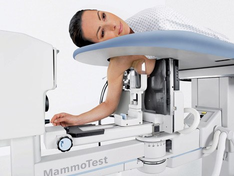
– In most cases, you will lie face down on a moveable exam table and the affected breast will be positioned into an opening in the table.

– The table is raised and the procedure is then performed beneath it. If the machine is an upright system, you may be seated in front of the stereotactic mammography unit.

– The breast is compressed and held in position throughout the procedure.

– Preliminary stereotactic mammogram images are taken.

– A local anesthetic will be injected into the breast to numb it.

– A very small nick is made in the skin at the site where the biopsy needle is to be inserted.


RadiologyInfo explains the pros and cons of this procedure:


Benefits:

– The procedure is less invasive than surgical biopsy, leaves little or no scarring and can be performed in less than an hour.

– Stereotactic breast biopsy is an excellent way to evaluate calcium deposits or masses that are not visible on ultrasound.

– Stereotactic core needle biopsy is a simple procedure that may be performed in an outpatient imaging center.

– Compared with open surgical biopsy, the procedure is about one-third the cost.

– Very little recovery time is required.

– Generally, the procedure is not painful.

– No breast defect remains and, unlike surgery, stereotactic needle biopsy does not distort the breast tissue and make it difficult to read future mammograms.

– Recovery time is brief and patients can soon resume their usual activities.

– No radiation remains in a patient’s body after an x-ray examination.

– X-rays usually have no side effects in the typical diagnostic range for this exam.


Risks:


– There is a risk of bleeding and forming a hematoma, or a collection of blood at the biopsy site. The risk, however, appears to be less than one percent of patients.

– An occasional patient has significant discomfort, which can be readily controlled by non-prescription pain medication.

– Any procedure where the skin is penetrated carries a risk of infection. The chance of infection requiring antibiotic treatment appears to be less than one in 1,000.

– Depending on the type of biopsy being performed or the design of the biopsy machine, a biopsy of tissue located deep within the breast carries a slight risk that the needle will pass through the chest wall, allowing air around the lung that could cause the lung to collapse. This is an extremely rare occurrence.

– There is always a slight chance of cancer from excessive exposure to radiation. However, the benefit of an accurate diagnosis far outweighs the risk.

– Women should always inform their physician or x-ray technologist if there is any possibility that they are pregnant. See the Safety page for more information about pregnancy and x-rays.


As medical imaging technology continues to advance, it is important to stay informed of the latest treatments so that your healthcare can become simpler and easier. Schedule your exam today and benefit!

CDI Miami | Tuesday December 15, 2015

FDA Approves Cap to Prevent Hair Loss During Chemo

Untitled design


Hair loss is one of the most despised side effects of chemotherapy, and new technology is now offering a way to save those precious locks.
Continue reading

CDI Miami | Thursday November 5, 2015

Optical Imaging Innovation Expected to Revolutionize Digital Imaging

Electrical engineers at the University of Wisconson-Madison have created the fastest, most responsive flexible silicon phototransistor ever made.

The phototransistor could improve the performance of myriad products – from digital cameras, night-vision goggles and surveillance systems to medical and digital imaging.

“Developed by UW-Madison collaborators Zhenqiang “Jack” Ma, professor of electrical and computer engineering, and research scientist Jung Hun Seo, the high-performance phototransistor far and away exceeds all previous flexible phototransistor parameters, including sensitivity and response time, according to the university.” – Security Sales

Similar to human eyes, phototransistors essentially sense and collect light, then convert that light into an electrical charge proportional to its intensity and wavelength. In the case of human eyes, the electrical impulses transmit the image to the brain.

“We actually can make the curve any shape we like to fit the optical system,” Ma says. “Currently, there’s no easy way to do that.”

One important aspect of the success of the new phototransistors is the researchers’ innovative “flip-transfer” fabrication method, in which their final step is to invert the finished phototransistor onto a plastic substrate. At that point, a reflective metal layer is on the bottom.

“In this structure – unlike other photodetectors – light absorption in an ultrathin silicon layer can be much more efficient because light is not blocked by any metal layers or other materials,” Ma says.

The researchers are patenting the technology through the Wisconsin Alumni Research Foundation.

Original article found here.

CDI Miami | Tuesday September 29, 2015

Chemotherapy May Not Be Needed for Early-Stage Breast Cancer

A recent study shows that early-stage tumors may respond to drug-therapy alone

Continue reading