


and how should I prepare?
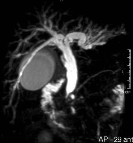
Image Credit: RadiologyInfo.org
Magnetic resonance cholangiopancreatography (MRCP) is a type of magnetic resonance imaging (MRI) exam that produces detailed images of the hepatobiliary and pancreatic systems, including the liver, gallbladder, bile ducts, pancreas and pancreatic duct. MRI’s use magnetic fields, radio frequency pulses and a computer to produce pictures of organs, soft tissues, bone and virtually all other internal body structures. However, MRIs do not use ionizing radiation (x-rays). These images help physicians determine pathology in the tissue layers, which can then be transferred electronically, or printed out.
When are MRCPs used?
- • Examine diseases of the liver, gallbladder, bile ducts, pancreas and pancreatic duct.
- • Help diagnose abdominal pain
- • Detect the underlying cause of pancreatitis
- • Provide a less invasive alternative to endoscopic retrograde cholangiopancreatography (ERCP).
Preparation Tips:
Before an examination, it’s customary to wear a loose-fitting garment such as a gown. Other precautions include avoiding food or drink at least a few hours before the procedure. MRI exams typically use a contrast agent as an effective part of the treatment. Your physician will ask if you have any allergies, as the contrasting material could potentially be an irritant. For pregnant women, MRIs are generally not advised as the magnetic field can negatively influence babies in the first trimester. If you are claustrophobic, ask your physician for a mild dose of a sedative to make you at ease for the procedure. All metallic objects need to be removed prior to the examination, as they can interfere with the magnetic field from the MRI.
These objects include:
- • Jewelry, credit cards, and hearing aids, all of which can be damaged
- • Pins, hairpins, metal zippers, and the like of which can distort MRI images
- • Removable dental work
- • Pens, pocket knives, and eyeglasses
- • Body piercings
Implanted biological devices (such as a pacemaker or implanted metallic joint prostheses) require a short time to settle after placement before it is safe to undergo an MRI examination.
Benefits vs Risks
Benefits:
- • MRI is a noninvasive imaging technique that does not involve the use of radiation
- • Magnetic resonance images scan the soft-tissue structures of the body, and the images provide more clarity than other imaging methods. This makes MRI invaluable in the early detection of cancer.
- • MRI is been proven effective in diagnosis of heart and vascular disease, stroke, and joint and musculoskeletal disorders.
- • MRI can help physicians evaluate how the organ is structured and how it works.
- • MRCP can produce images comparable to those obtained by a more invasive exam called endoscopic retrograde cholangiopancreatography (ERCP) without its associated risks including pancreatitis, or inflammation of the pancreas, perforation of pancreatic and bile ducts and bowel, and the risks for intravenous sedation required for ERCP.
Risks:
- • The MRI examination poses almost no risk to the average patient when appropriate precautions are taken.
- • Although the magnetic field is not harmful in and of itself, certain medical devices may malfunction or cause problems during an examination
- • In extreme cases, those suffering from kidney dysfunction can have a reactions to high does of the gadolinium-based contrast material used. Proper kidney assessment prior to treatment can effectively guarantee a successful treatment.
Although MRCP is a highly specialized form of an MRI, it has been proven effective in locating gallstones hiding away in the ducts surrounding the gallbladder. According to a recent study of 269 patients undergoing both ERCP and MRCP, the MRCP compared favorably next to the more invasive technique.
 "Why Do I Need an MRCP?")