


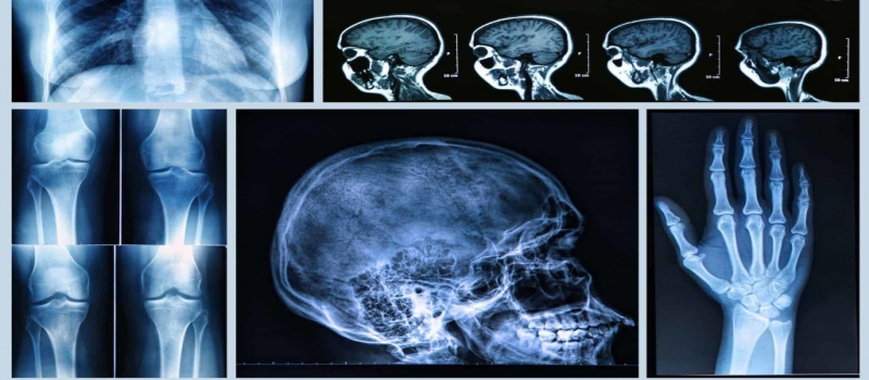
X-Ray is the most widely used imaging technique. X-Ray was invented in 1895 by Roentgen. X-ray Miami uses electromagnetic radiations to get a look beneath the skin of a person. With the advancement in technology, a clearer picture can be obtained which helps to see the problems in the skeleton system. X-rays penetrate soft tissues but not bones; that’s why the structure of the bones can be seen on an X-ray film.
How Do X-Rays Work?
To get a clear picture the patient is placed between an X-ray source and an X-ray detector. When the machine is turned on, X-rays go through the body and are absorbed in different amounts. Bones appear white on an X-ray in the black background of a radiograph.
What are the uses of X-rays?
- X-rays are used to examine bones for the possible fractures. It is a go-to tool to find out fractures of bones and teeth. X-rays help to find out the type of fracture; e.g. a hairline fracture, a broken bone, etc.
- X-rays are a useful tool in the dental world. Dentists advise X-rays before performing invasive dental procedures.
- X-rays also help to examine bone density; that is why they are done to find out if the patient is suffering from osteoporosis. Not only that, but X-ray is also helpful in detecting Arthritis.
- Chest X-ray is helpful to find out if the patient is suffering from pneumonia. Early imaging of pneumonia helps in saving a patient’s life.
- X-rays are extremely helpful in finding out any blockage in blood vessels and an enlarged heart.
- Abdomen X-rays are often advised to children who have swallowed any articles.
Mammography
Mammography is a technique which uses x-rays to find out any abnormalities in the breast tissues. Mammography is extremely helpful in finding cancer at an early stage and save the patient from chemotherapy and radiation therapy.
CT (Computed Tomography)
Computed Tomography is a technique which uses x-ray technology with computer processing to generate several cross-sectional images. Ct scan helps to get detailed images of bones and internal organs. These images can be generated in a 3-D plane.
Are there any risks involved?
X-ray is a handy technique which helps to see the internal structure of the body; it helps physicians to examine and plan a treatment plan according to the disease. Small exposure to x-ray doesn’t cause any life-threatening harm.
Pregnant women are not advised abdomen x-ray for the fear of radiation harming the fetus. X-ray to any other body part except the abdomen is regarded safe for the pregnant women.
Center for diagnostic imaging has expert radiologist which can help you in various imaging scans.
 "X-Ray Imaging Scan: Everything You Ought To Know")