Tag Archives: MRI

Magnetic resonance imaging (MRI) is a technique that uses a magnetic field and radio waves to create detailed images of the organs and tissues within your body.

CDI Miami | Tuesday January 5, 2016

New MRI-Guided Catheter Gives Hope for More Effective Stroke Treatment

Recent research at the University of California, San Francisco, has developed a tool that can navigate through blood vessels from the groin to the brain in search for blood clots using MRI-guided navigation. Known as the MARC catheter, it promises huge potential in the treatment of stroke and diseases under MRI guidance, and it is more efficient and expedient than its predecessors.


Because MRI is more useful than an x-ray when brain tissue is concerned, this steerable MRI-guided catheter could portend a huge development is stroke treatment.


According to UCSF, “The MARC – or magnetically assisted remove-controlled – catheter is based on a commercially available catheter, but made with nonmetallic fibers. There are microcoils embedded on the tip of catheter. By running a small electrical current a strong magnetic field on the tip of the catheter is created. That tip interacts with the strong magnetic field of the scanner, so we can steer through the body with remote control. The MRI guidance was done with a 1.5-tesla scanner.


This tool was easy to visualize under real-time MRI during the tests, as shown by research presented at last year’s Society of NeuroInterventional Surgery conference. Under magnetically assisted guidance, 192 of 240 turns (80 percent) were completed successfully, compared with 144 of 240 turns (60 percent) using standard x-ray guidance. Further, previous technologies for navigating under MRI are much slower rendering them more or less useless in clinical practice. The MARC catheter moved through blood vessels with a speed of 37 seconds per turn, compared to 55 seconds per turn of the conventional catheter- the manually directed catheter under MRI guidance.”

CDI Miami | Tuesday November 10, 2015

X-Rays and MRIs

Technology has come a long way in the X-Ray field as traditional film is largely being replaced by digital imaging, called digital X-Ray or digital radiography.  This new technology uses sensors from a digital image device as opposed to the use of traditional photographic film.  Widely used by doctors, dentists, hospitals, and X-Ray centers worldwide, this new technology allows for a more cost-effective and user-friendly approach to X-Ray imaging.

A key advantage to digital radiography is an increase in overall health and well-being.  By using digital imaging in place of traditional film, up to fifty percent less radiation is used to capture images.  In addition, when using traditional film, costly developing chemicals are required thus increasing chemical exposure to patients and staff members.  By removing the film-developing step from the X-Ray process, digital radiography provides a much safer approach and is more cost effective as well.  Both material and labor costs are lowered as developing chemicals are not required and companies that pick up hazardous waste are no longer needed, thus saving thousands of dollars.

Compared to traditional film, digital radiography produces higher quality images that are ready to view within seconds allowing for quicker appointment times. They are simpler to access and easily shared via email from practice to practice.  Similar to using photo edit controls on a computer, digital X-Ray images can be manipulated for clearer results by enlarging and brightening the images.  This provides a clearer picture, allowing for early detection and a more proactive treatment plan.  Because patients are also able to view the images more clearly, a new educational approach to diagnosis and treatment has been made possible.
04

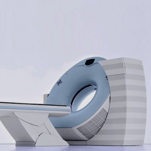
Also more and more patients and doctors are choosing open MRI scan vs. traditional MRI for their diagnostic needs. In fact, there is such a high demand that an increasing number of MRI diagnostic centers, such as the Center for Diagnostic Imaging in Miami, are offering open MRI either exclusively or as part of their diagnostic imaging MRI services.

Not only does open MRI offer the same high-quality diagnostic images as regular MRI machines, but it also offers a greatly increased comfort level for patients.

There are many groups of patients in particular that appreciate the openness of the open MRI tube, including children, overweight patients and those suffering from claustrophobia.

Open MRI has been revolutionary in being able to effectively scan children. And open MRI has not only allowed overweight patients a higher level of comfort, it produces better scans for this population. The open MRI’s wider opening allows a better positioning of overweight patients, which helps improve image quality and accuracy. The open MRI table weight limits are also substantially higher than those of closed MRI machines, providing access to a greater number of patients.

Many patients actually first discover that they are claustrophobic while getting a scan on a closed MRI machine. The closed tube, with limited space is extremely frightening for those that suffer from this phobia. Open MRI addresses these fears with its open sides and flared tube opening which allows a feeling of greater freedom and space.

Our Miami-based center offers the latest in all imaging technology: providing access to the latest treatments, while minimizing radiation exposure to our clients.

At the Center for Diagnostic Imaging, a full body scan can be offered in MRI scan form and also in CTA scan and Pet CT scan forms.

CDI Miami | Wednesday October 14, 2015

Saving Time and Money by Combining Diagnostic Medical Imaging Tests

Many patients do not ask questions about the types of diagnostic medical imaging tests their doctors order.  However, patients should not be afraid to start a dialog with their doctors and talk about whether it might make sense to have a second, different type of scan done at the same time.  In many cases, doing so can save the patient time and money.  Continue reading

CDI Miami | Tuesday September 22, 2015

5 Interesting Experiments Within MRI Scanners

Magnetic Resonance Imaging (MRI) machines are extremely sophisticated. They can detect tumors, see bone fractures, and examine electrical activity in the brain. You can conduct them in all sorts of positions: standing up, lying down, or when dancing a jig. It’s no surprise, then, that MRIs have been used for all sorts of off-the-wall experiments. Here’s some examples of the coolest types of MRI scanning:

  1. While Playing Jazz

Charles Limb, a hearing specialist at Johns Hopkins and a faulty member of the Peabody Conservatory, wanted to know hope some musicians are able to produce concert-length pieces of music that are entirely improvised, from beginning to end. So, he conducted MRI scans on the jazz pianists and rappers inside an MRI and had them perform. The imaging showed that the improvisers managed to shut off parts of their brains that handled self-monitoring.

  1. While reading T.S. Eliot

When an MRI scans an actor’s brain, it noticed a particular area of the brain – the infraparietal sulcus – light up. This area is important because it handles spatial memory.

A British researcher who was working on the project, commented: “I think actors’ brains are like musicians’ brains, in that they’ve been trained to learn enormous sections of language – not by rote, but by matters of association.”

  1. While giving birth

A woman in Germany gave birth in an MRI, giving viewers a rare glimpse into the inner-workings of the birth canal. Doctors could even watch the baby’s head change shape during the process.

  1. While playing video games

Researchers at the University of Illinois researched educational science by having test subjects play a game in various positions:

“We built in aspects of shifting your attention around to different objects in the display, remembering different pieces of information, using different rules depending upon the context and the changes in the game. There’s also a very complex psychomotor control in which you use a joystick to control a spaceship. We designed a somewhat entertaining task that was complex to learn, but built in various aspects of memory and decision making and control and attention. It was used very productively in studying learning and studying strategies that might be applied to enhance ideas about how we learn and how much and how quickly we learn.” – University of Illinois

  1. When introducing snakes and spiders

Bringing snakes and spiders in the room can definitely evoke the desired effect – fear.

“We are very grateful to Nachshon for inducing intense fear in the participants of our study,” deadpans Yadin Dudai, one of the lead researchers, in a video explaining the results.


CDI Miami | Tuesday July 21, 2015

MRIs and Peace – One Scan at a Time

At MIT, a collaboration is underway making the MRI scanner a new tool for resolving war and conflict. The goal of the lab is to use current knowledge of neuroscience to develop innovative, science-based conflict resolution strategies.

Continue reading

CDI Miami | Wednesday May 13, 2015

MRI’s and Tattoos: Possible Side-Effects

According to the FDA, more than 45 million Americans have a tattoo somewhere on their bodies. A tattoo is usually a form of self-expression, creates a fashion statement, or helps remember a special place or person. So, how do an MRI effect tattoos?
images

MRI’s have an effect on the iron within tattoos, as an MRI uses strong magnets along with radio frequency waves to gather data. Combined, they create an electromagnetic pulse that send signals from the MRI machine into our bodies. On the lighter side of the spectrum, tattoos can merely distort the images obtained from the scan. However, tattoos containing a high level of iron can heat and cause burns because of the magnetic field within the tattoo. During the imaging process, the current flowing can cause that metal within the iron to get hot enough to burn your skin. The inflammation can be anywhere from mild irritation to a second degree burn.

To make sure that you are safe, consult with your doctor prior to examination and discuss any concerns you may have. Your doctor will be able to evaluate the risks and provide suitable alternatives should a risk present itself. Some people are prohibited from having an MRI procedure done, especially if they have metal implants, such as cardiac pacemakers. Moreover, if you got your tattoo recently, such as the last 20 years, you won’t feel anything in the scan, as tattoo ink is metal-free. Previous to that time, tattoo artists were more prone to use ink that had metal filaments. These are the incidents that reported the negative side-effects from the scans, such as burns and blurred images.

Prior to getting an MRI, it’s important to remove all metal items you may have on you, such as: body rings, keys, paperclips or even hairpins. With magnetism, any metal object can quickly become a projectile missile. Not getting an MRI for fear of complications is much riskier than avoiding one, getting an MRI when you have a tattoo is safe as long as you take the necessary precautions as outlined above.

CDI Miami | Thursday May 7, 2015

National Cancer Institute grants $2.88 Million for Research and Development of Breast CT Scanning

CT scan of large breast tumor

CT Scan of a large breast tumor.
Image Source: RadiologyInfo.org

The National Cancer Institute recently awarded John M. Boone, a UC Davis professor of radiology and medical physicist, with an award of $2.88 million to advance and study computed tomography (CT) as a detection technique for breast cancer.


Continue reading

CDI Miami | Tuesday March 24, 2015

PET/MRI Better Than PET/CT for Foot Pain?

It’s true, according to a new study published in The Journal of Nuclear Medicince. This study evaluated 13 men and woman who had complaints of foot pain without a clear cause. The images from the PET scan combined with an MRI were significantly clearer than those combined with the CT scan. Continue reading

CDI Miami | Thursday December 4, 2014

Diagnostic Imaging Services for Arthritis

Arthritis includes more than 100 different rheumatic diseases and conditions, and it is one of the most common causes of disability in the country. According to the Centers for Disease Control and Prevention, approximately 50 million U.S. adults report having doctor-diagnosed arthritis. Continue reading

CDI Miami | Wednesday September 3, 2014

Can Sports Cause ALS?

Ever since the latest ice bucket challenge trend, ALS (Amyotrophic lateral sclerosis) has been all over the news. The attention has also awaken a prolonged scientific debate about whether exercise or pesticides might somehow contribute to the development of the fatal neurodegenerative disease.

Ever since the great Yankees first baseman Lou Gehrig died of ALS in 1941 at age 37, many Americans have vaguely connected A.L.S. with athletes and sports. However, it remains unclear whether exercise is indeed a risk factor and what types of exercise may be of concern. In Europe, the possible connection to sports has been more popularly discussed. In the past decade, a few widely publicized studies indicated that professional Italian soccer players were disproportionately prone to ALS. But these studies were extremely small and had methodological problems. So to better determine what role sports and exercise might play in the risk for A.L.S., major new studies would have to be combined.

One idea that researchers offer is that soldiers on active duty are engaged in strenuous physical labor. Or, they are exposed to toxins which could play a role in ALS. The Association is helping to fund an effort by the Veterans Administration to collect information about veterans diagnosed with ALS, to shed light on possible environmental factors associated with the disease. This VA registry will also serve as a stepping stone to clinical trials. Both possibilities, of toxin exposure or the influence of intense exertion, are advanced when researchers consider the finding that Italian soccer players appear to have increased incidence of ALS.

For now, he and other scientists are continuing to study Italian soccer players, as well as athletes in other sports, including rugby, which, for some reason, confers no increased risk of A.L.S., although it involves considerable contact. Such research may ultimately shed some light on the underlying mechanisms of the disease, which are still poorly understood.

About Center for Diagnostic Imaging – For over a decade the Center for Diagnostic Imaging Miami has constantly pushed towards the future of medicine so that we may provide the most optimized care for their patients.

We truly appreciate our patients and strive to meet their needs. We are always ready to make you our number one priority. For more information call us at 1.800.371.0002 or visit our Contact page.