


 Nuclear medicine, ionizing radiation and radiopharmaceuticals are all terms that patients hear when their doctors order diagnostic imaging tests such as X-rays, CT scans, PET scans and bone scans.
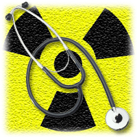
Nuclear medicine uses radioactive substances in the diagnosis and treatment of disease. These substances can include ionizing radiation – from a PET or X-ray machine for example – or radiopharmaceuticals, which are injected into the body and emit ionizing radiation.
A good example of nuclear medicine is a cardiac stress test, which can help doctors determine if parts of the heart are not healthy. During a nuclear stress test, a small amount of a radiopharmaceutical is injected into the patient. A radiologist can then use a specialized camera to see where rays are being emitted from inside the body. A computer uses those signals to generate a clear picture of the heart and where there are problems with decreased blood flow.
With all this discussion of radioactive materials, some patients naturally become nervous about what impact these tests will have on future health. Nuclear medicine has been linked with a very small, but increased cancer risk in some patients, which leads some people to believe that getting the test in the first place may not be such a good idea.
However, ionizing radiation gives doctors useful information that cannot be obtained from other types of tests, like ultrasounds. If the test is done at a reputable diagnostic imaging center, the dose of radiation will be the lowest possible amount required to obtain a clear picture of the area in question. Those clear images help doctors develop life-saving treatment plans that well offset the risk of increased radiation exposure.
For example, the American Cancer Society and FDA estimate that a patient’s exposure to 10 mSv (millisievert, a measurement of radiation dose) from an imaging test may increase the risk of death from cancer by about one chance in 2,000. To put that into perspective, the average person is exposed to 2 mSv a year from the natural environment. A typical chest X-ray generates a radiation dose of 0.1 mSv, and a heart CT scan is about 16 mSv. Even with higher-dose tests, cancers from diagnostic imaging usually take many years to develop. It’s far more likely a patient will die of causes unrelated to nuclear medicine.
That said, patients should always ask their doctors and radiologists questions so they understand why the radiation is necessary, what the risks are, whether there are any viable alternatives, and what benefits they will gain from having the test. Patients can then rest assured that the benefits are greater than the small increased risk of cancer from radiation exposure.
Should Patients Fear Radiation Exposure from Diagnostic Imaging?
| Share |
 "Should Patients Fear Radiation Exposure from Diagnostic Imaging?")
|
Tags: Nuclear Medicine