


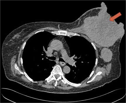
CT Scan of a large breast tumor.
Image Source: RadiologyInfo.org
The National Cancer Institute recently awarded John M. Boone, a UC Davis professor of radiology and medical physicist, with an award of $2.88 million to advance and study computed tomography (CT) as a detection technique for breast cancer.
Boone has pioneered breast CT, having worked with a large team of scientists and physicians over the past 15 years to develop the technology. His team has built four breast CT scanners, each with successive improvements in spatial resolution.
The research, called “Breast CT: Final Steps to Translation,” will involve 400 women who have been recognized to have suspicious lesions requiring breast biopsy with traditional methods.
The new project will make two comparisons. In the first study, Breast CT will be compared with mammography to establish which treatment is better at detecting lesions in the breast tissue. The second study will focus on comparing contrast-enhanced magnetic resonance imaging (MRI), with contrast-enhanced breast CT. I contrast-enhanced imaging, the patient is injected with a contrast agent, which helps distinguish vascular tumors, such as those in the breast.
The aim of the study is to determine if breast CT is better at detecting potentially malignant lesions.
“Because breast cancer can present as both soft tissue masses and as micro-calcifications, this means that our newest scanner needs to detect both better than digital mammography,” he said in a recent news release. “Only then will breast CT technology be able to improve breast cancer detection rates in women at normal risk for breast cancer.”
The goal of the second comparative analysis is to demonstrate the effectiveness of breast CT with contrast compared to MRI, and if the technique takes less time for both the patient and the radiologist diagnostic assessment.
“If shown to be equivalent to contrast-enhanced breast MRI, contrast-enhanced breast CT would be a viable and far more cost-effective tool for imaging women with suspicious lesions,” Boone said. “This would likely reduce the negative biopsy rate and increase the positive predictive value of breast imaging in general.”
Currently at UC Davis, more than 300 patients underwent breast CT. In a partnership with the University of Pittsburgh the technique has scanned further 300 patients. The research team has published nearly 50 papers on breast CT in the past 15 years.
“Our long-term goals are to show with our newest scanner that we can improve cancer detection rates in the screening population over that of mammography and tomosynthesis,” Boone said. “By increasing the performance of breast cancer screening in a practical and cost-efficient manner, we hope to move the standard-of-care to true 3D breast imaging in order to improve care and increase survival in women with breast cancer, and importantly to also reduce over-treatment of women with benign findings.”
 "National Cancer Institute grants $2.88 Million for Research and Development of Breast CT Scanning")