


Receiving a Heart MRI in Miami is a useful way to diagnose, treat and monitor cardiac conditions
An MRI, short for magnetic resonance imaging, is used to create detailed images of the organs and tissues within the body, including the heart. Sometimes referred to as a heart MRI or a cardiac MRI, this procedure is usually performed when a doctor needs to diagnose or monitor cardiac disease or surgical changes in a patient. Unlike a CT scan, an MRI does not use radiation. It is considered a safer alternative for pregnant women.
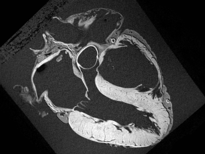
MRI of a human heart
A cardiac MRI in Miami allows doctors see how various parts of the heart are functioning, including its chambers, valves, major vessels and surrounding structures. It can be used to gather more information for symptoms related to:
- • Aneurysm – dilation in part of the heart muscle or the aorta.
- • Atherosclerosis – the gradual clogging of the arteries.
- • Cardiac tumor – a tumor located on the outside of the heart, or within one of its chambers or muscle tissues.
- • Cardiomyopathy – enlargement of the heart.
- • Congenital heart disease – defects of the heart which occur in utero.
- • Congestive heart failure – the inefficient pumping of blood caused by a weakened heart.
- • Valvular heart disease – failure of the heart valves which can obstruct blood flow within the heart.
As with any Miami MRI procedure, a cardiac MRI uses a magnetic field and radio waves. This creates detailed images of the heart that can help doctors evaluate any number of cardiac conditions and plan the correct course of treatment. When undergoing a cardiac MRI in Miami, patients can expect that the test will be carried out by a technician who is specifically trained in MRI technology. The results are interpreted by a radiologist, who reports the findings to the patient’s physician.
One of the challenging aspects of a cardiac MRI is that the heart is constantly in motion. MRI machines generate the clearest pictures when the area being imaged is perfectly still. Therefore, special techniques are used during cardiac MRIs that are not as common with other types of tests. For example, the MRI may be synchronized with electrocardiography (ECG) tracing, or the patient may be asked to breathe a certain way during the procedure in order to improve the image quality.
As with any diagnostic imaging test, follow-up exams may be required if there is a suspicious or unclear result. A series of cardiac MRIs may also be ordered to determine the effectiveness of treatments.
A cardiac MRI does not use radiation and is considered to be a safe, non-invasive procedure. However, due to the magnet, some patients may not be good candidates for the exam. This includes anyone with certain implanted devices like pacemakers or drug infusion pumps, and those with some types of prosthetic devices or intrauterine contraceptives. Other metal objects like bullets, shrapnel, surgical clips, pins, plates, screws or wire mesh can exclude patients from having an MRI in Miami or elsewhere.
There may be additional risks based on a patient’s medical history and current health. Patients are encouraged to ask their doctors and MRI technicians questions, and to be completely honest when filling out pre-procedure paperwork. Giving doctors all of the facts up front can help mitigate any risks associated with the test, so patients won’t have anything to worry about.
 "The ‘Ins & Outs’ of a Cardiac MRI Procedure")