Tag Archives: diagnostic medical imaging

CDI Miami | Wednesday April 3, 2019

A Brief Overview Of Positron Emission Tomography

A Brief Overview Of Positron Emission Tomography


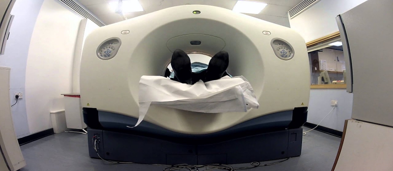
Positron emission tomography is a nuclear medicine imaging system done using small amounts of radioactive material. Diagnostic medical imaging Miami centers are extensively using this method to detect and find the severity of heart disease, cancer and other abnormalities in the human body.
 
Positron emission tomography used to treat serious diseases like endocrine, gastrointestinal, and neurological disorders as nuclear medicine procedures can pinpoint every tiny molecular activity in the human body. The potential of PET to recognize disease from the initial stages has helped the doctors and patients significantly.
 
The noninvasive nature of nuclear medicine imaging has made it the most common diagnostic method to evaluate medical conditions.
 
What Is A Radiotracer?
 
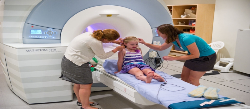
The radiotracer is the radioactive material used in imaging scans with the intravenous injections. The procedure of PET starts with radiotracers used within small quantities to be detected on the PET scan. It is widely used to bind particular proteins in the human body. They are specifically considered for accumulating cancerous tumors or sections of inflammation within the body. F-18 fluorodeoxyglucose also called FDG is the commonly used radiotracer and is a very similar molecule to glucose.
 
The cancer cells are more metabolically active that absorbs glucose at a higher rate in the body. The PET scans detect the higher rate quickly as compared to other diagnostic medical imaging scans, allowing the doctor to find the disease early. FDG is amongst the few widely used radiotracers for detecting the range of conditions in the body.
 
Common uses of PET or CT scan Miami
 
This procedure is used to sense cancer and then find how it has spread in the body. It is used to assess the extent of the effect of various therapies in the human body and make the physicians aware of any harmful effects on the body. The doctors change the treatment plan after assessing the side effects on the body after cancer therapy. Moreover, it is very helpful to determine whether, after the treatment, cancer has returned.
 
It is frequently used to check the blood flow to the heart muscle and other surrounded area. PET is also used to find the effects of myocardial infarction and heart attack on various areas of the heart. In addition to this, it is proficient to map normal heart and human brain function. These scans are widely used to recognize areas of the heart affected after some abnormalities or disorders in the body.

CDI Miami | Wednesday March 20, 2019

How PACS Transformed The Medical Imaging?

How PACS Transformed The Medical Imaging?

PACS or picture archiving communications systems has changed the scenarios of radiology departments drastically. With a number of PACS system installed in the radiology departments of the hospital, the diagnostic medical imaging departments have progressed enormously in the country. With this system, there are a number of advantages to the medical professionals, as they can capture, stock, send and display the medical images from one radiology workstation to another workstation and can use the images for future research.

Impact Of PACS

PACS has completely changed the working of the hospital’s radiology department. PACS has made scheduling easy, the patient can track and generate reports after some time in that system. Patients X-ray and CT-scan images are electronically stored in the system, which can be viewed afterward on the computer screen by the doctors for treatment and research purposes. PACS has improved the diagnosis method and creates a film in fewer images. The radiologist or doctor has the full access to view the past images of the patients in center for diagnostic imaging for comparing multiple images through PACS.

PACS has enhanced the working of the radiology department. It has improved the productivity of diagnosis and reporting of particular diagnostic imaging. Hospital and healthcare centers are giving best services to the people coming for various diagnostic tests. Moreover, PACS have completely simplified the hospital’s process, as they can operate by viewing various X ray radiology images.

The prominent feature of PACS is the speed, which has solved the matter of the time-consuming process of images developing. But now images can be seen in PACS after few seconds.

Importance Of PACS In Radiological Department

The PACS system has made diagnostic imaging more in digital forms which are more affordable and has resulted in finishing the possibility of losing any images due to mismanagement by the authorities. In addition to this, the diagnostic methods have improved giving a clearer image to the doctors. Doctors can manipulate the images for correctly diagnosing and getting a full detailed image on the screen.

The feature of PACS to be accessible to multiple clinicians has helped patients to show their reports to various doctors. Additionally, they are being used in training the interns and enhancing the teaching experience for a better understanding of the medical students. Now, medical students are exposed to various diagnostic digital images for having in-depth knowledge of the web. PACS is easy to use and highly effective method adopted by hospitals which have resulted in improving the working and diagnosing process of various departments.


CDI Miami | Thursday February 28, 2019

Breast Cancer-Overview And Symptoms

Breast Cancer-Overview And Symptoms

Sharp pain or tenderness in the breast can be something serious which you have never imagined. A breast lump is the first thing that makes women visit a doctor or center for diagnostic imaging. Though breast cancer usually displays no symptoms in the initial stage, if it is detected on time, it could be successfully treated. Whenever women have pain or tenderness in their breast, they suppose it to be breast cancer. Conversely, breast pain is hardly the first clear symptom of breast cancer.
 
There are many other factors that can cause breast pain which is clinically termed as mastalgia. The main cause of this pain is either changeability of hormones due to menstruation or when women have birth control pills or take infertility treatments. Moreover, when the bra doesn’t fit well or have large breasts, they usually have a neck, back or shoulder pain. Women having bra cyst or who are extremely stressed at times get a breast pain which does not mean its breast cancer.
 
Breast Screening
 
Breast screening discovers breast cancers early by using an X-ray test called 3d mammogram to spot cancers which are too less to feel or see. However, due to some risks of breast cancer screening, one should be aware of.
 
The chances of getting breast cancer rise with age, every woman from 50 to 71 years should get breast cancer screening every 3 years. If you’re concerned about breast cancer indications, like a lump or thickened tissue in a breast, or breasts look or feel different than normal, don’t wait for screening, see a general practitioner as soon as possible.
 
There are some women who have a genetic tendency, family history, or certain factors which indicate breast cancer. They should be screened with MRIs and mammograms, to detect it from an early stage. It is suggested to talk with a clinician about the risk for breast cancer and the appropriate screening plan for it.
 
CHECK With Your Doctor If
 
If something unusual is suspected in your breast, check with your doctor or nurse as soon as possible. It’s imperative to keep checking it even if you have regular diagnostic medical imaging screening.  As these symptoms don’t essentially confirm breast cancer, but they are also the cause of other ailments about which you should be worried. In case your doctor asks for further testing, or refer a breast clinic specialist, you should get yourself checked at the earliest.

CDI Miami | Monday February 25, 2019

Positron Emission Tomography- Purpose And Risks Involved

Positron Emission Tomography- Purpose And Risks Involved


Positron emission tomography (PET) is the latest imaging technology for assessing your tissues and organs functioning at the cellular level. PET CT of Miami recognizes the changes at the cellular level, to find the early onset of disease before it’s detectable on other imaging tests.

It comprises of radiotracers, a special camera, and a computer to evaluate your organs and tissue function. It starts with the injection of a short-acting radioactive substance, called radiotracer that is soaked by biologically active cells. After that, the person is placed in a tunnel-like device which detects and translates the emitted radiation into three-dimensional images.

The images help in recognizing the abnormalities in the metabolism of a cell and tissues in the human body. The PET scan diagnoses and assesses the brutality of a wide range of diseases that can take place in the body. Various serious diseases such as cancer, heart disease, and disorders of the brain can be detected from the initial stages with the help of diagnostic imaging Miami.

Purpose of test

Positron emission tomography has many diagnostic applications but is generally done when the doctor either suspects cancer or want to know how much cancer has spread. Adding to this, it is used to evaluate the status of the patient’s heart before the bypass surgery, when other imaging tests are inadequate. It is always done when Alzheimer’s disease is suspected or to see the status of the brain while treating refractory seizures.

Moreover, it is used to see the extent of damage done after the heart stroke or stroke to seek the proper treatment. It is also used to monitor the response of your body to various cardiovascular, neurological, or cancer treatments, aiding in making the treatment more safe and swift.

Risks Involved In PET

A PET scan poses few risks and is a painless diagnostic medical imaging procedure because the quantity of radiotracer used while imaging is so less that there is no need to follow standard radiation precautions. The radiotracer is glucose with a radioactive isotope attached, whose drug half-life is enormously short. In most cases, the radiotracer is in and out of your system within a day.

The injection causes localized pain and swelling and at times allergic reactions. The most significant risk in a few cases is claustrophobia; you should inform your doctor about this before as being placed inside the tube-like device makes people nervous. In some extreme cases, the doctor may prescribe a mild sedative, to help reduce anxiety during PET.

CDI Miami | Thursday February 14, 2019

The vital importance of CT Scan in diagnosing the underlying problems

The vital importance of CT Scan in diagnosing the underlying problems


CT scan is one of the top five diagnostic medical imaging procedures as it has proved to be very successful in diagnosing deadly diseases which would otherwise cannot be detected with the x-rays. Both CT scan and x-rays are conducted to take internal images of the body, however, in case of x-rays the internal body parts overlap and therefore does not give detailed and clear images of the organs.
 
Diagnosing a certain disease or condition is difficult with x-rays but with the advent of CT scan technology, even a tiny part can be scrutinized clearly as the organs does not overlap each other and the scan offers a clear image of the body part.
 
When there’s a need to go for a CT scan?
 
Particularly, a CT scan is needed to examine people who might be suffering from internal injuries caused due to car accidents or due to various kinds of trauma. Doctors usually suggest for CT scan when they want visualize any organ that might be causing abnormal symptoms in the body or if they want to plan any medical or surgical treatment for a certain disease diagnosed.
 
There are certain reasons for why a CT scan Miami is required which includes diagnosing muscle and bone disorders such as tumors and fractures, detecting the location of the tumor or blood clot, monitoring and detecting stage of cancer, heart disease, lung and liver diseases, checking the effect of treatments carried out, identifying internal injuries, and it also helps the doctors in performing various procedures such as surgery or radiation therapy.
 
Certain kinds of risks associated with CT scan
 
It gets more detailed images of the organs, so you might be exposed to more radiation than x-ray radiology. However, the low doses used in the scan have not led to any long-term harm but if the doses are high, there might be risk of cancer. But the results and the benefits of scan outweigh the small potential risks. Doctors always prefer to use least possible dose to acquire the needed information.
 
You must share any medical condition with your doctor before undergoing a CT scan for example- if you are pregnant it might be dangerous for you. Although CT scan might not affect your baby, the doctor will suggest you to go for ultrasound or MRI in order to save your baby from even small amount of radiation.

CDI Miami | Monday February 11, 2019

MRI- a better diagnostic tool for detecting the underlying problems

MRI- a better diagnostic tool for detecting the underlying problems

With the evolving technological era, medical diagnostic tools and procedures are also taking a new way to deal with the issues inside the body. Nowadays, there are numerous procedures that help the doctors diagnose the diseases early and give them a chance to cure them on time.

Some of the procedures include MRI, CT-Scans, X-rays, PET-Scans etc. However, MRI is one of the most reliable diagnostic medical imaging procedures that use magnetic field and radio waves to see through the body.

If you are facing any bone issues, X-rays are good at it as it takes images of the calcium inside the body, however, MRI Scan images water which is more useful as the tissues inside the body are made up of water. So this helps the radiologist to take high-resolution images of various organs and tissues which might not be possible with the X-ray.

When it’s important to go for an MRI?

An MRI Scan is useful when there’s a need to diagnose the conditions that affects the soft tissues inside the body. This might include detecting tumors in various parts of the body, damaged ligaments or injuries in the soft tissues, a joint disease or injury, spinal injury or diagnosing the internal organs such as brain, heart, and digestive system.

Thus, MRI Scan Miami have proven to be very successful in providing the detailed images of the soft tissues inside but in case of bone tissues they are not much effective. This is the reason why bone injuries are always investigated with the X-rays.

MRI Scan is a safe procedure however the complications may arise such as if you have worn some metal objects during the scan, it might cause injury, sometimes, the magnetic field might damage the internal heart pacemaker or in other case the patient might be allergic to the contrast dye injected to get a clearer image of the body parts.

Share any medical conditions before undergoing an MRI Scan

v
You should tell your doctor in case you have any metal implants inside your body as they may get damaged with the magnetic rays. Well the affects of MRI on fetus are unknown, however, if you are pregnant you must tell it to your doctor. The other factors that need to be considered include fasting before getting an MRI Miami, which means you will be advised not to drink or eat at least 4 to 5 hrs before undergoing the Scan.

Also, tell your doctor if you are claustrophobic so in that case the doctor may give you some medication to make you relax during the procedure.

CDI Miami | Wednesday February 6, 2019

PET Scan- a more sophisticated technology to diagnose the diseases and treat them effectively

PET Scan- a more sophisticated technology to diagnose the diseases and treat them effectively

PET scans are usually recommended by the doctors when they need to diagnose or devise the treatment options for a number of medical conditions. The doctor will provide you with the detailed description that why this diagnostic medical imaging is essential for your treatment.
 
This technology known as positron emission tomography lets the doctor take the images of your body and see through the organs in order to check any underlying diseases that might be causing abnormal symptoms.
 
PET scan uses a unique dye which contains radioactive tracers that are injected into the body through a vein in your arm depending upon the type of body part that is going to be examined.
 

Why a doctor will recommend you the one?

 
There are certain reasons why a doctor will suggest you to get a PET scan Miami which includes diagnosis of the type and the location of cancer, they consider this scan more reliable as they have the potential to differentiate between the benign and malignant cells. With the help of this scan the doctor can identify if the symptoms are associated with cancer or not.
 
The radiologists might use the PET scan to carry out an effective Neurological assessment in the brain. Various forms of dementia can be determined along with diagnosing the recurrence of cancer in the brain.
 
In case you have a coronary heart disease or you have recently suffered from cardiac arrest, the doctor can suggest you a cardiac PET scan. This will assist the doctor in cardiac bypass surgery. So in all cases, PET scan is a more efficient and reliable way to diagnose and treat the diseases.
 

Risks related to PET scan and what precautions to follow

 
As you know the PET scan make use of radioactive tracers which might sound to you somewhat risky however, exposure to the radiation is very less. The amount of radiation in the tracers is low so your body is at a minimal risk. But if you are still worried you can ask the doctor about the possible risks it can cause.
 
So it would be recommended that you should go for PET ct of Miami if you feel any abnormal symptoms occurring in your body. The benefits associated with the test outweigh the risks involved and the test can prove to be beneficial in the long run. However, people with certain allergies and medical conditions should alert their doctor before undergoing the scan.

CDI Miami | Monday January 28, 2019

Reasons to Rely only on a Prominent Imaging Scanning Service Provider

Reasons to Rely only on a Prominent Imaging Scanning Service Provider

There could be several reasons for which your physician may advise an imaging scan to analyze the cause of any ailment. It is always a wise decision to seek help from the professionals that are experienced enough in delivering the best in class imaging scan services that can help in getting the desired test results without any hassle.

There are numerous options when it comes to choosing the best in class diagnostic medical imaging service provider that can harmonize your medical scanning needs. Here is a list of reasons why you should always consider visiting a renowned professional when it comes to getting adequate medical imaging scan services.

Precise Results and no chance of Variation

One of the biggest issues that most of the people face is the variation in the test results that eventually affects their overall medical treatment. Therefore, it is necessarily important for an individual to rely on a service provider that can offer the finest services that ensure adequate preciseness in the test results without any glitches.

As per the experts, there is no point you can rely on the ones that are hardly experienced in delivering the best in class medical imaging services. This is the reason you have to do some research regarding the imaging diagnostic center in Miami service provider so as to get the assurance of quality services. Moreover, you also get accurate results without worrying about any variation.

Quick Delivery of Accurate Reports
You may be aware regarding the fact that a little delay in the submission of the reports to your physician could be quite risky. Thus; you have to be sure enough to rely on the ones that hold a great reputation in the market. There are people who usually ignore the importance of a reputed medical imaging service provider that can lend a hand in augmenting your medical treatment.

You get the assurance of quick delivery of the reports when you rely on any of the renowned medical imaging service providers in Miami. You can also download the reports online. All you need to do is to get some references regarding the same from your acquaintances that have earlier been to any of these service providers so as to get the reliable services. You can always explore the internet for the same where you would be getting detailed information about the same.

CDI Miami | Thursday December 13, 2018

Chronic heart conditions can now be detected through CT scans

Chronic heart conditions can now be detected through CT scans
 
CT scans are now used to examine the chronic heart conditions, the physician may recommend for a CT scan to examine the anatomy of the heart, the coronary circulation, and different vessels if an abnormal symptom is experienced.
 
CT scan Miami helps detect the underlying conditions that are causing abnormalities in the body, these conditions might include heart disease, tumors, clots, and other defects. Sometimes coronary diseases are not determined in a CT scan so you must keep in mind that things like a heart attack cannot be predicted completely.
 
What you should expect during a Cardiac CT Scan?
 
Initially, when you will visit the hospital, the nurse will record your weight, height and B.P. your blood may be taken for a lipid profile test. The technologist will make the patient to lie on a scanning table, he will place small sticky electrodes on the chest, these electrodes are attached to the electrocardiograph which monitors your heart’s activity and produces it in the graphical form.
 
During the CT scan, you will be placed inside a doughnut-shaped scanner, then the machine will take several images of your coronary system to examine the various areas of your heart.
 
The diagnostic medical imaging is done by the cardiovascular radiologist who analyzes the images to look for any calcification in the arteries. If there isn’t any calcification the exam is considered to be negative.
 
The doctor will prohibit you to take caffeine 24 hours before the test. You are supposed to drink only clear liquids before the day of the examination.
 
What are the symptoms of coronary heart disease?
 
Pain in the chest: The patient may feel tightness or pain in the chest which is usually referred to as angina. It is generally triggered by physical and emotional stress. The pain lasts for several minutes, in some individuals the pain may reach up to the neck, arm or back.
 
The patient may experience shortness of breath as the heart may not be able to pump the blood properly to meet the needs of the body. It can also cause extreme fatigue to the patient.
 
In case the patient has complete blockage of the coronary artery, he or she can get a cardiac arrest or heart attack. The early signs of heart attack include chest pain, shortness of breath or sweating. Sometimes it can occur without any warning signs or symptoms.
 
If you experience any warning signs you must go for PET CT in Miami in which the radiologists use radioactive material to get information about various functions of the body like blood flow and glucose metabolism. It helps in diagnosing the heart disease more efficiently sometimes.

CDI Miami | Wednesday December 12, 2018

Medical imaging- a key to patient healthcare

Medical imaging- a key to patient healthcare
 
Medical imaging is extremely important as it helps in the prevention of many chronic diseases such as cancers and tumors. The doctors can detect, diagnose, and start the treatment as early as possible to prevent the patient from fatal diseases.
 
Diagnostic medical imaging uses a number of non-invasive methods that allows the radiologists and other medical personnel to see through the body and diagnose the underlying problem. Various organs and areas inside the body can be seen and treated with the help of medical imaging. It plays a crucial role in the healthcare of the patient.
 
Why medical imaging is one of the top developments in the face of clinical medicine?
 
It literally saves the life of a patient: The advancement in medical imaging has enhanced the screening of diseases and helps in the early diagnosis of acute and chronic conditions. In the absence of medical imaging, many of the severe and life-threatening diseases may go unnoticed until they reach a much more fatal stage.
 
Medical imaging has reduced the requirement for intrusive exploratory surgery: The development of technologies such as Magnetic Resonance imaging (MRI), Computed Tomography Scan (CT scan), and Echocardiography, x-ray services etc. has dramatically reduced the need for surgeries.
 
A huge amount of money is saved: With the right scan at the right time, the patient is able to get the proper treatment, thus costs as well as time is saved. As it replaces the need of surgery and also decreases the amount of time the patient has to remain in the hospital, so less cost is incurred for the treatment as well.
 
The doctors are able to identify the exact underlying problem that is causing the abnormal symptoms in the body and are able to give the proper treatment based on the reports of the patient.
 
Which tests involve ionizing radiation that might have a harmful effect?
 
There are generally four types of technologies that are used in medical imaging namely x-rays, MRI, Ultrasound, and nuclear medicine. The patient will be exposed to radiation depending upon the type of test carried out.
 
X-rays, 3d mammography, and fluoroscopy give out lesser radiation than the CT scans. The MRI and ultrasound do not emit any radiations as these can harm the children who are more sensitive to the radiation. The risks involved in medical imaging are less.
 
However, you should go for the imaging that is required to examine the exact condition. The benefits provided by medical imaging outweigh the risks associated with it.