Tag Archives: diagnostic center

CDI Miami | Thursday February 21, 2019

Why Do You Need A 3d Mammography?

Why Do You Need A 3d Mammography


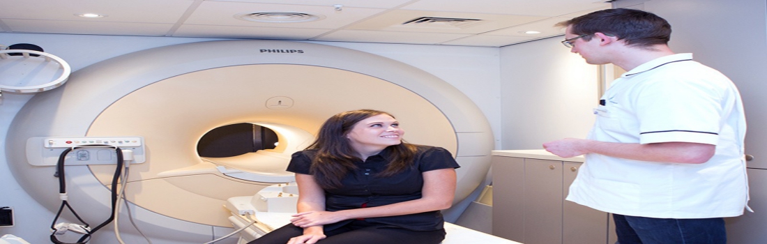
Early detection of any cancer increases the probabilities of recovery by successfully treating it. Every woman in her mid-30s should get an annual mammogram for early detection of breast cancer.
 
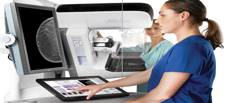
A 3D mammogram (breast tomosynthesis) is the latest imaging test that combines multiple breast X-rays (like traditional mammography) for creating a three-dimensional image of the breast tissues for detecting any signs or symptoms of breast cancer or any other abnormalities. It is used to examine the reason for various breast problems, such as lumps, nipple discharge, and pain. 3D mammography produces highly detailed images of breast tissue by 3D capture of multiple slices of the breast from different angles.
 
The 3D mammogram takes approximately 300 images, giving an exact idea of the issue whereas two-dimensional mammogram takes about four images at a time giving some false alarms. It lets the radiologist view the breast tissue in full detail, equipping them to give a certain diagnosis, if they have cancer or not by visiting the nearest diagnostic center. This is specifically valuable for women with dense breast tissue, as they are at higher cancer risk.
 
Why is 3D Mammography Performed?
 
We know that traditional mammography took only two images of each breast of a side-to-side view and a top-to-bottom view. 3D mammography takes various X-ray images of the breasts from multiple angles to create a digital version of internal breast tissue. The multiple images make radiologists assess the breast in 1mm slices in place of only two views.
 
Radiologists correctly interpret results from 3D mammography in dense breast tissue, resulting in fewer false-positive and false-negative readings.
 
What to Expect from 3D Mammography
 
The preparation of a 3D mammography is exactly the same for traditional mammography. Women should schedule mammography at a date one week after their menstrual period starts because they are least tender at that time.
 
3D mammography involves a bare torso, so it is suggested to wear comfortable, easy-to-remove clothes. Avoid applying Deodorant, perfume, lotion, and other products on underarm or torso before 3D mammography, since these products interfere with the imaging process. Women should inform their radiologist or physician if they are pregnant, breastfeeding, have breast implants or suffer from any medical conditions, so they can change the procedure accordingly.
 
A radiologist interprets the results of 3D mammography by viewing the calcification or masses in the breast tissue and using a standardized classification system called Breast Imaging Reporting and Data System (BI-RADS).

CDI Miami | Wednesday February 20, 2019

How has diagnostic imaging impacted medical science?

How has diagnostic imaging impacted medical science


Diagnostic imaging, commonly stated as radiology, is one of the most revolutionary innovations in the medical world which has changed the way physicians and patients view health and diseases. The biggest advantage of the latest technologies, instruments, and equipment has led to substantial improvements in the world of medicine reducing the treatment cost and time period of the treatment by completely identifying the ailment from the images at diagnostic center.
 
Radiology has made medical professionals, to look in the living human body without cutting it to detect & treat the life-threatening diseases and other complications in the patient. It is the visual discipline of the medical community and is a central component for the growth and development of medical science as a whole. Moreover, without radiology, it is impossible to recognize the contemporary medical fields which are wholly based on treatment after diagnostic imaging.
 
Here are a few points on how the advances in medical imaging technology have changed medical science.
 
Upgraded monitoring and diagnosis for medical professionals
 
Before the development of modern medical imaging technologies, almost every medical diagnosis required invasive procedures which harmed the human body. However, the development of x ray services, MRI and ultrasound technology has made medical professionals across the globe to take better care of patients. The discovery of medical imaging is highly instrumental in the success and development of medicine.
 
It is helping medical professionals to see medical conditions better; dynamically helping the treatment of medical conditions. X-ray imaging and gamma radiation are amongst the most prevailing medical imaging technologies for active treatment.
 
Picture Archiving and Communications System (PACS)
 
Picture Archiving and Communications System acronym or PACS is a technological solution to make it easy to store and access the vast volume of medical images in the radiology department. Medical professionals utilize a PACS for the wide management of images in medical imaging and to use it for reference to treatment.
 
Moreover, digital access to the updated version of a patient’s medical images, clinical reports, and history helps in the better treatment and improve the care for a quick recovery, reducing the possibility of treatment and prescription errors & stopping redundant testing, saving a lot of expenses and resources of the patient and doctor. Digital access improves patient safety and saves both healthcare facility expenses and patient time and money. The system stores, retrieves, and share images produced by various medical hardware modalities in the diagnostic center for women Miami, such as from an X-ray, ultrasound machines, computed tomography (CT) scan, magnetic resonance imaging (MRI) and many more.

CDI Miami | Thursday January 3, 2019

Guidelines to help you detect and treat breast cancer

Guidelines to help you detect and treat breast cancer

Go for a breast cancer screening
Before a woman gets any signs or symptoms of breast cancer, screening is important to diagnose any abnormalities and start its treatment at an early stage. Although the screening cannot prevent breast cancer, it can surely detect it early and you can get started with the proper medication and therapy.

Consult with your doctor about which screenings are suitable for you and at what age you should have them. Nowadays, a 3D mammogram is a new and advanced technology used to detect breast cancers with more accuracy and efficiency.

Initially, self-awareness is important to judge any symptoms of cancer. You must be familiar with the look and feel of your breasts. If you feel any changes in the size of your breasts or feel any lumps or pain you must consult your doctor soon in order to clarify your doubts.

Tests conducted for breast cancer screening

Mammograms are highly recommended for detecting breast cancer at an early stage when it is easier to treat. Going for regular mammograms reduces the risk of dying from cancer.

MRI of breasts is also conducted to detect cancer. It uses magnetic and radio waves to take images of the breast. This test is used along with the diagnostic mammogram for women who are at high risk of having breast cancer.

A clinical breast exam is used by the doctors to feel the lumps or other changes in the breast, it is usually not reliable and the patient is suggested to go for the screening tests such as mammograms.

Risk factors you can change to prevent breast cancer

The women need to be physically active or otherwise, they might be at the risk of getting cancer. Having overweight after menopause can also pose the risk of breast cancer. Women with normal weight are at less risk of getting cancer, so it is crucial to maintaining a good weight in order to avoid any risks.

Women who take hormone replacement therapy including estrogen or progesterone can increase the risk of breast cancer. In addition to this, oral contraceptives such as birth control pills also tend to raise the risk of cancer.

Having pregnancy after the age of 30 or not breastfeeding can increase the risk of breast cancer.

In any case, if you feel any abnormal symptoms in your breast, you must visit the diagnostic center near you to clear your doubts and start your treatment early not to land up in a serious stage.

CDI Miami | Monday December 10, 2018

Importance of a CT Scan – for the early detection of a major underlying problem

Importance of a CT Scan- for the early detection of a major underlying problem
 
CT scan is more than an ordinary X-ray as a combined set of beams are simultaneously sent through the body to get the images from different angles with more clarity. These images are then examined by the doctors to get an insight into any underlying problem inside the body.
 
At the diagnostic center, you will be placed inside a doughnut-shaped machine and it is less likely that you will feel claustrophobia.
 
What are the most common uses of CT Scans?
 
Initially, CT Scans were used to take the images of the brain but as the technology has become more advanced, these are being used to take pictures of nearly every part of the body. The CT Scans offer cross-sectional and highly detailed images of the organs so they are extensively used by the doctors to examine various parts of the body such as chest, abdomen, and pelvis etc.
 
These are highly recommended as doctors can see through the body without operating or performing any difficult examinations.CT Scans are used for a variety of reasons, some of them are:
 
Doctors use CT scan for detecting different kinds of cancers such as lung, kidney or liver.
 
If the patient has suffered from serious injuries, it is used to check for any inner injuries or deformations.
 
Sudden onset of symptoms such as abdominal pain, breathlessness etc. may let the doctor recommend a CT scan.
 
Doctors use CT scans for the preparation of radiotherapy as it will be easy for them to know where exactly a tumor lies inside the body.
 
CT scans are also used to detect vascular diseases and skeletal problems inside the body.
 
These are particularly used to diagnose the brain tumors and test for bleeding in the brain.
 
The imaging center Miami are well-equipped with high-end and advanced machines to provide you with sophisticated scans and accurate results and diagnosis.
 
Are CT scans dangerous? Do they hurt?
 
The amount of radiation involved in the scan is small. However, you are exposed to a beam of X-rays rather than a single x-ray so doctors only recommend a CT scan only when it is highly necessary.
 
For pregnant women, it is usually not suggested as the radiations can harm the fetus. It is only recommended when there is serious condition involved.
 
It does not hurt anyway but some people just get anxious while lying inside the scanner. Make sure you are calm and cool and just tell your doctor if you are feeling nervous or panicky.
 
CT scan Miami has experienced staff and has years of expertise, thus they provide full support and perform the scan carefully without harming the patient.

CDI Miami | Monday December 3, 2018

Facts about breast cancer-is it a matter of concern?

Facts about breast cancer-is it a matter of concern?

One of the top concerns in women nowadays is breast cancer. Nationalized breast screening programs, center for diagnostic imaging, modern treatment methodologies, along with the research going on the disease, all these play an important part in the detection and prevention of breast cancer.
 
Risk factors associated with developing breast cancer
 
If you have a family history of breast cancer that is if your sister, mother or daughter had a breast cancer, you might have the risk of developing cancer in your breast.
 
Age factor also plays a crucial role in having this type of cancer, as you get older, the risk of developing cancer also increases. The women between 55 to 59 years of age have are at higher risk of having this cancer. While it primarily affects older women but it can also occur in women under the age of 30.
 
If you earlier had cancer in one of your breasts, this might increase the risk of having cancer in the other part of the similar breast or in the other breast.
 
Overweight and alcohol intake also increases the risk of  cancer. You must go to the breast diagnostic center in case you feel lumps in your breasts as it could possibly be due to the breast cancer.
 
In case if you have a family history of other cancers such as cancers of ovaries, uterus, colon or cervix, this can increase the risk of breast cancer.
 
If you have undergone a hormone replacement therapy, you can have the risk of cancer due to the prolonged use of oestrogen and progesterone.
 
If you have given birth to a child after 35 years of age or you have never given the birth to a child can also increase the risk of having cancer.
 
How can you reduce the risk of having a breast cancer?
 
• Limit the consumption of alcohol and avoid smoking: Alcohol and smoking can apparently increase the risk of breast cancer so doctors recommend avoiding these two to prevent you from cancer.
 
• Try to maintain a proper weight: If you are overweight especially after menopause, you are at higher risk of developing cancer so try to control your weight and have a balanced diet.
 
• Regular exercise can prevent you from breast cancer: 75 minutes of vigorous exercise weekly can help lower the risk of cancer.
 
• It would be best to visit the women center Miami to get proper guidance and treatment regarding the cancer and its prevention.

CDI Miami | Thursday November 1, 2018

How to Prepare yourself for an MRI Scan

How to Prepare yourself for an MRI Scan

It’s perfectly fine to get worried and feel nervous if you never had an MRI scan before, but there’s no need to fear the MRI scan. It’s a painless and a completely safe process and will not harm you in any case. Here’s a guide to give you an insight of the MRI scan so that you should feel less stressful and can confidently go to the medical imaging center.
 
Principles of MRI Scan
 
MRI-Magnetic Resonance Imaging uses powerful magnetic fields, magnetic field gradients, and radio waves to take the images of your internal organs, and tissues. The doctors usually conduct this test to diagnose the underlying problem causing the abnormal symptoms in your body. The imaging center Miami does not use radiations like X-rays or CT scans.
 
Get ready to undergo an MRI scan!
 
1. Tell your doctor if you have claustrophobia- fear of confined places: As during the MRI scan you will be enclosed in a tube-like machine, so if you suffer from claustrophobia, you will be given the sedatives before the test in order to prevent you from the panic attack.
 
2. Inform the doctor about any health issues: Tell your doctor if you have any of the following conditions:
• Diabetes
• Kidney problems
• Pregnancy
• Allergic to iodine or gadolinium
 
3. Follow the instructions given by your doctor before the exam: Generally you will continue with your regular routine but if you have any medical conditions then the doctor may give you some guidelines that you must follow.
 
4. Don’t go alone for the exam, ask a friend or family member to accompany you: It will be good to take someone with you to the diagnostic center as the process is lengthy and quite stressful. It will make you feel better.
 
5. Remove any items that contain metal: You need to arrive early as there’ll be some paperwork and the nurse may need to talk to you before the scan. The radiologist may refrain you from eating about an hour before.
 
You need to remove all the jewelry, hairpins, watches, wigs before going into the machine.
 
6. Relax and Relax: The noise of the clicking of the magnets and the fear of closed in a tube can make you anxious. So just take some deep breaths and close your eyes before being placed into the scanner.

CDI Miami | Thursday August 30, 2018

How to Locate the Best Diagnostic Center for Women Miami for Reliable Services

diagnostic center for women Miami

Seeking the prominent diagnostic center for women Miami? Well, in that case, you have landed at the right location! Many people hardly emphasize the overall reputation of a diagnostic center when it comes to imaging scans. It is mandatory for you to check the background of a service provider that can assure quality services without any hassle!

There’s no point in spending your money on the services that are actually not going to serve you in the treatment of a specific disease. A lot of people face issues with their reports like poor quality imaging, delayed results, and uncertainty. One should always keep in mind that they can always get the right treatment from a renowned physician only when they get a proper diagnosis as recommended by their doctor.

Thus; it is necessarily important for you to pick the right diagnostic center for better results and timely reports. Dive in here to know more about choosing the appropriate service provider.

Always Research on the Internet

It is always a wise decision to rely on the reviews posted by people who have earlier been to any of the diagnosis centers located in Miami. This would not only give you an idea regarding the services offered by a particular center but you could eventually decide which would be best for you.

Another reason to rely on these online portals is that you can get the idea about the services that are sometimes not available at all the diagnostic centers. Once you have shortlisted the available service providers, it is the time to pick the one that suits you the most.

Seek References

Undoubtedly, you can always rely on references from your acquaintances that possess some knowledge about the promising diagnostic medical imaging service providers in your location. A referral could be the best thing that can surely lend a hand in finding the appropriate service provider possessing great expertise in offering the best in class imaging diagnosis services.

Apart from this, you can always seek references from your physician as they possess ample knowledge about the promising centers in Miami. It would be great to consider them for your scans like the X-Ray, Mammogram, or an MRI scan.

So these are some of the easiest ways to seek professional medical imaging services that can augment your overall medical treatment.