Tag Archives: 3D mammography

Officially tomosynthesis, a revolutionary screening and diagnostic tool designed for early detection of breast cancer that can be done in conjunction with a traditional 2D digital mammogram.

CDI Miami | Tuesday August 2, 2016

Preventative Steps, Risk Factors, and Screenings for Breast Cancer

A cancer is named based on where it first develops. Breast cancer begins in the breast tissue, eventually forming a mass of cancerous cells called a tumor. Cells from that tumor can break away and spread (called metastasis) to other organs of the body – such as the lymph nodes, bones, lungs, and even liver—where they can multiply in number.


Even though the causes of breast cancer are not known, early detection and newer treatments are leading researchers toward a cure. Researchers have identified certain risk factors that have been associated with breast cancer. Knowing these and determining an individual’s risk are the first steps toward breast cancer protection.


It is almost impossible to predict whether or not a woman will be diagnosed with breast cancer – some women can have all the risk factors and never develop the disease while other women develop the disease and do not have many of the known risk factors. As with all diseases, there are certain factors that place some women at higher chance of developing breast cancer than others. Some risk factors can be controlled while others cannot.


Breast Cancer Action Early Detection Saves Lives Logo

logo credit: Breast Cancer Action

Certain steps can be taken to reduce the risk of breast or other cancers. Cancer experts recommend eating a balanced diet that includes plenty of fruits and vegetables and is low in saturated fats. For those who consume alcohol on a daily basis, they should speak with their doctor about personal risks for breast cancer and heart health. Avoid weight gain, particularly after menopause and exercise to maintain a healthy weight.


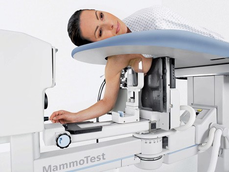
While getting an annual mammogram is crucial, 10 to 15 percent of breast cancers are not detected by the test and additional breast cancers may also become detectable between routine mammograms. That is why it is important for every woman to perform her own breast self-exam every month in addition to getting a yearly mammogram or clinical breast exam. The best time for a breast self-exam is roughly seven days after the beginning of a monthly menstrual cycle, because this is when the hormone levels from the ovaries are at their lowest and breasts are least likely to be tender or swollen. Women who have gone through menopause should examine their breasts the same day each month, such as on the first day of the month.


Screening refers to tests and exams used to find a disease in people who do not have any symptoms. Breast cancers found during screening exams are more likely to be smaller and still confined to the breast. The size of a breast cancer and how far it has spread are some of the most important factors in predicting the prognosis of a woman with this disease.


The Center for Diagnostic Imaging and the Comprehensive Breast Care Centers specialize in the early detection of breast cancer. The fully trained staff and board certified radiologists utilize the best medical imaging solutions available today, such as breast ultrasound, breast MRI, and 3D mammography. They insure that each exam is tailored to each patient’s individual needs.

CDI Miami | Tuesday April 12, 2016

The Economic Value of 3D Breast Tomosynthesis

Breast cancer is the most common cancer among women. Despite increasing incident rates, annual mortality rates have declined by approximately 2% per year, which has been attributed to the combination of earlier detection and improvements in treatments.

Mammography plays a key role in early breast cancer detection. Two-dimensional digital mammography is the current standard of care in breast cancer screening. An abnormal finding with screening mammography prompts additional imaging with diagnostic mammography, targeted breast ultrasound or breast magnetic resonance imaging. These additional studies may determine the need for a breast biopsy in order to determine if the area of concern is benign or malignant. A considerable percentage of women to be called back for additional imaging or biopsy do not ultimately have cancer (i.e. a false positive test result) resulting in unnecessary patient anxiety and economic burden. Continue reading

CDI Miami | Wednesday January 20, 2016

Stereotactic Breast Biopsy

Stereotactic breast biopsy uses mammography – a specific kind of low-dose breast imaging that helps locate a breast lump or abnormality and remove tissue sample for examination under a microscope. It’s less invasive than surgical biopsy, leaves little to no scarring and can be an excellent way to evaluate calcium deposits or tiny masses that are not visible on ultrasound.

What is Mammographically Guided Breast Biopsy?

A breast biopsy is performed to remove suspicious area in the breast and examine them under a microscope. Suspicious areas may include:

    • An unidentified mass
    • Microcalcifications, a tiny cluster of small calcium deposits
    • A distortion in the structure of the breast tissue
    • An area of abnormal tissue change
    • A new mass or area of calcium deposits is present at a previous surgery site


How should I prepare?

Before the exam, you may be asked to remove some or all of your clothes and to wear a gown during the exam. You will have to remove jewelry, dental appliances, eye glasses and any metal objects or clothing that might interfere with the x-ray images.
You should also report all medications that you are taking – including herbal supplements.

How is the procedure performed?

– Breast biopsies are usually done on an outpatient basis.

– In most cases, you will lie face down on a moveable exam table and the affected breast will be positioned into an opening in the table.

– The table is raised and the procedure is then performed beneath it. If the machine is an upright system, you may be seated in front of the stereotactic mammography unit.

– The breast is compressed and held in position throughout the procedure.

– Preliminary stereotactic mammogram images are taken.

– A local anesthetic will be injected into the breast to numb it.

– A very small nick is made in the skin at the site where the biopsy needle is to be inserted.


RadiologyInfo explains the pros and cons of this procedure:


Benefits:

– The procedure is less invasive than surgical biopsy, leaves little or no scarring and can be performed in less than an hour.

– Stereotactic breast biopsy is an excellent way to evaluate calcium deposits or masses that are not visible on ultrasound.

– Stereotactic core needle biopsy is a simple procedure that may be performed in an outpatient imaging center.

– Compared with open surgical biopsy, the procedure is about one-third the cost.

– Very little recovery time is required.

– Generally, the procedure is not painful.

– No breast defect remains and, unlike surgery, stereotactic needle biopsy does not distort the breast tissue and make it difficult to read future mammograms.

– Recovery time is brief and patients can soon resume their usual activities.

– No radiation remains in a patient’s body after an x-ray examination.

– X-rays usually have no side effects in the typical diagnostic range for this exam.


Risks:


– There is a risk of bleeding and forming a hematoma, or a collection of blood at the biopsy site. The risk, however, appears to be less than one percent of patients.

– An occasional patient has significant discomfort, which can be readily controlled by non-prescription pain medication.

– Any procedure where the skin is penetrated carries a risk of infection. The chance of infection requiring antibiotic treatment appears to be less than one in 1,000.

– Depending on the type of biopsy being performed or the design of the biopsy machine, a biopsy of tissue located deep within the breast carries a slight risk that the needle will pass through the chest wall, allowing air around the lung that could cause the lung to collapse. This is an extremely rare occurrence.

– There is always a slight chance of cancer from excessive exposure to radiation. However, the benefit of an accurate diagnosis far outweighs the risk.

– Women should always inform their physician or x-ray technologist if there is any possibility that they are pregnant. See the Safety page for more information about pregnancy and x-rays.


As medical imaging technology continues to advance, it is important to stay informed of the latest treatments so that your healthcare can become simpler and easier. Schedule your exam today and benefit!

CDI Miami | Tuesday December 22, 2015

3D Mammography – 21st Century Breastcare

CBCC


If caught early, breast cancer can be treated with a very high success rate. Breast cancer is one of the most treatable cancers because of the advancements in lumpectomy and mastectomy surgery, and the early detection methods that have been developed in recent times.
Nevertheless, the discovery of any type of cancer is extremely distressing. It has a huge impact on people’s lives. Breast cancer is particularly upsetting for women, because their breasts are considered part of their femininity and sexuality.


The Center for Diagnostic Imaging always recommends that women have regular mammograms so that their physicians can detect any occurrence of cancerous cells at a very early stage. This is the stage at which non-invasive treatments can usually get the cancer sufferer into remission. In order to have the scans done, a woman must find a convenient imaging center that complies with her physician’s standards. Many women in South Florida choose one of the CDI Miami offices, conveniently located in Aventura, North Miami Beach and South Dade.


The Center for Diagnostic Imaging always recommends that women have regular mammograms so that their physicians can detect any occurrence of cancerous cells at a very early stage. This is the stage at which non-invasive treatments can usually get the cancer sufferer into remission. In order to have the scans done, a woman must find a convenient imaging center that complies with her physician’s standards. Many women in South Florida choose one of the CDI Miami offices, conveniently located in Aventura, North Miami Beach and South Dade.


Medical imaging can get expensive, so it is vitally important that patients find a center they can trust, where they know they will receive the best quality images that do not contain any errors.The South Florida CDI offices have the very latest imaging equipment available in every center. Fully qualified radiologists, experts at using the scanners and reading the results, are employed at every CDI location.The Center for Diagnostic Imaging’s facilities in North Miami Beach uses 3D mammography, an imaging technique that can detect breast cancer at a very early stage. An MRI scan can pinpoint disease centers in the body and these can be monitored on a regular basis for any changes, which could indicate future potential problems. People searching for a pet scan in Miami will find that CDI Miami comes highly recommended, based on the local popularity of the centers and the high quality of service offered at every location. CDI Miami even gives patients the option to have a full body CT scan. This will give the radiologists an entire map of what is going on inside the body so that potential disease can be detected. This information is then passed onto the patient’s physician. Any abnormalities that show up can then be treated immediately, or monitored to check for any developments. So many people who end up with life-threatening illnesses and conditions could have been saved a lot of pain and anguish if they had been able to detect problems and treat them at a very early stage.


About CDI Miami: CDI Miami is the Center for Diagnostic Imaging, the premier center for CT scan Miami with locations in South Florida at Aventura, North Miami Beach, and South Dade. For more information and to book a consultation, call toll free on 800-371-0002 or visit cdimiami.com


CDI Miami | Tuesday December 8, 2015

3D Mammography Aids in Early Cancer Detection

According to a new study, digital breast tomosynthesis, or 3D mammography, can improve the detection of breast cancer, according to a new study.


Continue reading

CDI Miami | Wednesday October 14, 2015

Saving Time and Money by Combining Diagnostic Medical Imaging Tests

Many patients do not ask questions about the types of diagnostic medical imaging tests their doctors order.  However, patients should not be afraid to start a dialog with their doctors and talk about whether it might make sense to have a second, different type of scan done at the same time.  In many cases, doing so can save the patient time and money.  Continue reading

CDI Miami | Tuesday September 29, 2015

Chemotherapy May Not Be Needed for Early-Stage Breast Cancer

A recent study shows that early-stage tumors may respond to drug-therapy alone

Continue reading

CDI Miami | Friday May 23, 2014

Breast Ultrasound Technology

Breast ultrasounds can assist in the diagnostic evaluation of breast tissue and abnormalities and is often used to assist in distinguishing between fluid and solid masses. Ultrasound imaging, also called ultrasound scanning or sonography, involves exposing a part of the body to high-frequency sound waves to produce pictures of the inside of the body. Ultrasound exams do not use ionizing radiation, which are used in X-rays. Because ultrasound images are captured in real-time, they can show the structure and movement of the body’s internal organs, as well as the blood flowing through the blood vessels.

Ultrasound Miami imaging is a non-invasive medical test that helps physicians diagnose and treat medical conditions. Ultrasound imaging of the breast produces a picture of the internal structure of the breast. The Doppler method is a special ultrasound technique that calculates the blood flowing through blood vessels, including the body’s major arteries and veins in the abdomen, arms, legs, and neck.

During a breast ultrasound, the radiologist may use this Doppler technique to evaluate the blood flowing (or the lack of blood flowing) into any breast mass. In some cases, this provides information as to the cause of suspicious masses in the breast.

Breast ultrasounds are mainly used to help determine and diagnose breast masses and abnormalities found during physical exams. Ultrasound imaging will help to determine if a mass is solid and cancerous or cystic and benign. In some cases, ultrasounds help to provide additional information about breast abnormalities.

Ultrasounds can even substitute for mammography imaging in some cases because ultrasounds can detect small abnormalities that may not be visible with regular mammography or even 3D mammograms. This kind of ultrasound screenings are especially helpful for women with dense breasts, silicone breast implants, very little breast tissue, and those who are at a high risk for breast cancer.

In an ultrasound examination, a transducer sends the ultra-sound waves and records the echoing waves. When the transducer is pressed against the skin, it directs small pulses of inaudible, high-frequency sound waves into the body. As the sound waves bounce off of internal organs, fluids and tissues, the sensitive microphone in the transducer records tiny changes in the sound’s pitch and direction. These signature waves are instantly measured and displayed by a computer, which in turn creates a real-time picture on a monitor. Frames of the moving pictures are typically captured as still images.

Another kind of ultrasound, the Doppler ultrasound, measures the direction and speed of blood cells as they move through vessels. The movement of blood cells causes a change in pitch of the reflected sound waves (called the Doppler Effect.) A computer collects and processes the sounds and creates graphs or color pictures that represent the flow of blood through the blood vessels.

Ultrasounds are also useful for biopsy procedures. An ultrasound-guided biopsy requires no exposure to radiation as a radiologist uses sound waves to locate the suspicious breast mass. This procedure is very useful when suspicious changes can’t be detected with the use of a mammogram. This type of biopsy is a minimally invasive way to obtain a sample of breast tissue for further diagnosis, as well as a faster and less painful option than a traditional surgical breast stereotactic biopsy.

The Center for Diagnostic Imaging and the Comprehensive Breast Care Centers specialize in the early detection of breast cancer. They utilize the best digital medical imaging solutions available, to best help the needs of each individual.

CDI Miami | Friday January 10, 2014

CBCC Announces More Options for Surgical Breast Lump Removal

The Comprehensive Breast Care Center, a preferred breast MRI guided biopsy Miami facility, announces additional procedures to remove harden lumps discovered during routine breast examinations.

When a suspicious lump is discovered during an examination, physicians usually request additional tests to determine if the lump should be removed. If numerous test results cannot conclude the lump is harmless, the physical will schedule a biopsy.

A breast biopsy is a procedure which removes suspicious breast growth.
Depending upon how big the lump is, the breast MRI Miami clinic will determine the type of biopsy which needs to perform. The two types of breast lump removal biopsies: surgical and core-needle.

A Surgical biopsy is where the surgeon makes a one- to two-inch cut in the breast, removing suspicious tissue.
During a core-needle biopsy, the surgeon removes small amounts of harden tissue through a hollow needle inserted into the breast.
Once the biopsy is completed, the extracted tissue will be examined by a pathologist to determine if the lump contains any signs of cancerous cells. Once the examination is complete, the pathologist will forward the results to the patient’s family physician.

Although mammograms may not be an enjoyable experience, they are necessary in early detection of breast cancer.
Physicians recommend annual checkups to ensure women remain healthy. Earlier detection increases a woman’s odds of being treated and increasing the chance for survival.

About CBCC, Miami:
The Comprehensive Breast Care Center prides itself on being an accomplished and dedicated imaging center for Breast care and analysis in South Florida. They specialize in early detection of breast cancer, provided by a fully trained staff and team of Board Certified Radiologists.
Utilizing the best medical imaging solutions available today, CBCC ensure that each exam is tailored to a patient’s individual needs.
The CBCC center keeps in strict accordance with the patient’s physician’s instructions. Since they analyze the exams themselves they are able to provide prompt results and deliver them to the physician within 24 hours.

Located in the city of Aventura, in North Miami and South Dade, the CBCC is easily accessible from all mayor roads and highways, making it the ideal center digital mammography center for anyone in the Miami/Fort Lauderdale area.

The Comprehensive Breast Care Center currently offers the following scanning services:
–       Digital mammogram
–       Breast MRI/Ultrasound
–       Breast Biopsies
–       Bone Density
–       Genetic Testing
–       Breast Navigator
–       Complete Breast Care Program

CDI Miami | Monday September 9, 2013

Using the Best Technology for Diagnostic Imaging

Diagnostic imaging is saving lives every day. By detecting the presence of malignancy early on, before a tumor is developed, cancer sufferers can be treated and make a full recovery from certain types of cancer. There is new research into the treatment of cancer being conducted all the time. There are frequent breakthroughs in medical research, and there are more and more types of cancer that are becoming treatable. The success stories and personal accounts from cancer survivors are always very encouraging. This is great news for anyone who has a family history of certain types of cancer, and who are therefore most vulnerable to developing the disease. With early detection, and the most innovative treatment programs, even people at high risk have a good chance of staying cancer free. However, the detection of cancer requires top quality imaging equipment.

CDI, the Center for Diagnostic Imaging, one of the top diagnostic facilities offering breast MRI guided biopsy in Miami, announces new assurances that the very best technology is being used for the early detection of cancer. People searching for breast ultrasound in Miami will find that CDI appears at the top of local listings. The Center for Diagnostic Imaging in Miami has the best ultrasound equipment available. An ultrasound can show vital information about any abnormalities in the body, by using high frequency sound waves to produce images. CDI are also a top choice for people searching for PET scan in Miami, again because the scanning equipment is the best available. A PET scan delivers important information about abnormalities in the body. By having their patients ingest a small amount of radioactive material and then photographing the whole body using a scanner, physicians can gain a lot of information about any problem areas and conduct more tests, such as biopsies. CDI also specializes in 3D mammography. Before any cancer treatment is undertaken, it is important to get as much information as possible about what is going on inside the body. Sometimes there is more than one affected area or organ, and physicians have to take this into account before deciding on a course of treatment. Offering the most advanced technology used for cancer detection, CDI Miami is a “one stop shop” for physicians to send patients to for tests. Each type of test reveals different information and these results can be combined to give the clearest diagnostic picture possible.

About CDI Miami: CDI Miami, the Center for Diagnostic Imaging, is often the number one choice for people searching for MRI in Aventura and surrounding areas. There are convenient locations in South Florida – Aventura, North Miami Beach and South Dade. For more information and to book a consultation, call toll free on 800-371-0002 or visit cdimiami.com

bestmritechnology